Apoptosis opens urodeal membrane, cavitates coprodeum,
and removes chick urogenital ducts
S.A. Miller, C. Clark, and E. Crary
Department of Biology, Hamilton College, Clinton, NY 13323
Abstract
American Zoologist 37:162a (1997)
Morphogenesis of chick cloaca involves cavitation of occluded
cords of cells and removal of membranes and certain urogential ducts.
Chick cloaca is described as three successive chambers that are
distinct around midgestation, but become indistinct in adults. At 9
days of incubation two of these chambers, urodeum and proctodeum, are
separated by urodeal membrane, whereas the coprodeum has not yet
cavitated from rectal cord. Between day 9 and 10 the membrane
ruptures, and occluded cord begins to cavitate. To test the
possibility that programmed cell death (PCD) known as apoptosis is
involved, we applied the immunomarker ApopTag™
(Oncor®) to sections of chick embryos representing 12 hour
intervals of development between days 8 and 10. Abundant and dense
collections of apoptotic cells in day 9 membrane and in rectal cord
suggest that PCD is important in morphogenesis of cloaca and caudal
gut. Apoptosis was also heavily marked in adjacent urogenital ducts.
Apoptosis removes tail gut and remnant of the primitive streak
(Miller and Briglin, 1996) in earlier
stages. Programmed cell death is clearly a major contributor to
morphogenesis in the caudal axis of chick embryos.
Supported by Hamilton College Academic
Fund for Seniors. Includes Senior Theses of Clark and
Crary.
|

|

|
|
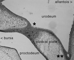
Urodeal membrane (cloacal plate) in a 9-day chick embryo
shows dark spots of apoptosis (*) and early formation
of cell cords (**).
|
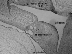
Urodeal membrane (*cloacal plate) in a
9.5-day chick embryo shows dark spots of apoptosis and
established cell cords. Apoptotic markers in some cells of
the rectal cord (**) suggest the beginning of
cavitation of the coprodeum.
|
related information on apoptosis in remodeling of chick embryo
cloaca and hindgut
to SAMiller's research
to SAMiller's publications to SAMiller
's homepage


Last Modified: 14 November
1999


![]()
![]()