Apoptosis and the Development of Avian
Integument:
Feathers and Periderm
Christopher Reamer and Sue Ann Miller, Department of Biology,
Hamilton College, Clinton, New York
Abstract
Illustrations
below
Apoptosis, also known as programmed cell death or PCD, is a
specific form of non-random cell death that is an integral part of
vertebrate development. PCD is involved with mammalian and avian
digital development and epidermal cell differentiation in chick
embryos (Carlson, 1999), but the potential role of apoptosis in
development of the avian integument has not been examined. We
investigated the role of apoptosis in early feather morphogenesis and
shedding of the late-stage periderm in chick embryos.
We examined the feather development and periderm sloughing
processes by employing an in situ immunomarker for apoptosis
(ApopTag®, Intergen).
Feather and skin tissue sections from White Leghorn chicken embryos
(SPAFAS) at 14, 16, and 18 days of incubation were studied.
Apoptotic cells were marked in feather sections from chicks at all
three ages. Positive markers were observed in the distal end of
14-day feathers. 16-day feathers showed light staining which was
localized in the mid-axial region of developing feathers. Sections of
18-day feathers showed positively marked apoptotic cells in the
feather follicle. Apoptotic bodies within feathers were observed
mainly in the thin layer of epithelium bounding the medulla of the
ramus within individual barb ridges. Apoptotic cells within the
feather were distributed along the longitudinal axis of the feather,
and localized distal to proximal with increasing age. Periderm
sections of 14- and 16-day chicks showed no marked cells, whereas
periderm sections from 18-day chicks showed abundant and very clearly
marked apoptotic cells.
This preliminary investigation supports our hypothesis that
apoptosis plays a role in both sculpting of feathers and shedding of
the periderm. As is the case with cell differentiation within the
feather, apoptosis seems to move proximally along the longitudinal
axis of the feather as development progresses. Furthermore, there is
strong evidence to suggest that the loss of the periderm is
temporally specific, and does not occur until day 18 of incubation.
This finding is in agreement with earlier observations of timing of
periderm loss (Bellairs and Osmond, 1998). Apoptosis appears to be
the mechanism by which the periderm is removed.
Grant sponsor: Sigma Xi Grant-in-Aid of Research;
Grant sponsor: The Hamilton College Academic Fund for Seniors.
Chris presented this research as a poster at "Celebrating Student
Research at the Millennium", a Sigma Xi Student Research Symposium
held at St. Joseph's University in Philadelphia on 28
April 2000. His travel to present his work was funded by the Biology
Department Student Travel Fund. A full manuscript is in preparation
as earlier stages are currently being studied to provide a more
complete understanding of patterns of cell death and documentation of
cell division.
Apoptosis in
Feather Sections
Brown nuclei mark apoptosis
in all figures.
|
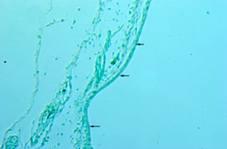
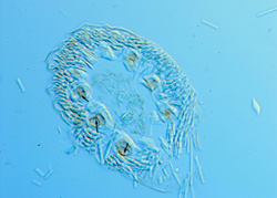
14-day
distal feather section: Strong positive markers are
localized in the distal tips of 14-day feathers and are
bound to the outside of the medulla of the ramus. Proximal
sections of a feather bud do not mark for apoptosis.
Marginal and axial plates have already been removed by day
14 of incubation.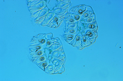
|
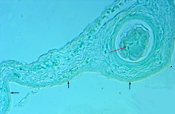
 16-day
mid-axial feather section: Positive markers
surround the medulla of the ramus with a mid-axial
localization. Feathers have begun to keratinize by day 16 of
incubation, which makes obtaining sections
difficult. 16-day
mid-axial feather section: Positive markers
surround the medulla of the ramus with a mid-axial
localization. Feathers have begun to keratinize by day 16 of
incubation, which makes obtaining sections
difficult.
|
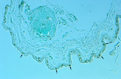
16-day
[A] and 18-day [B] follicle sections:
Sections of 18-day feather follicle mark for apoptosis
where there were no markers at 16 days. Undifferentiated
calamus of the feather within the follicles contains many
positively stained nuclei that appear to occur randomly
within the follicle.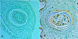
|
Apoptosis in Periderm Sections
Black arrows indicate the
periderm layer.
to research on earlier
feather germs
to SAMiller's research
page
to SAMiller's publications
to SAMiller
's homepage


Created 1 May 2000 Last
Modified: 11 August 2003