Apoptosis Plays A Role in Removal of the
Thyroglossal Duct and in Remodeling of the Thyroid Rudiment of Chick
Embryos.
Sue Ann Miller, Samuel J. Klempner, Christine Campbell and
Elizabeth Ransom
Department of Biology, Hamilton College, Clinton, New York
Abstract
Extensive modification is an important part of avian
pharyngenesis. Aortic arch arteries form and are then removed or
altered, pharyngeal pouches are remodeled as their closing plates
rupture, and the thyroglossal duct is lost after the thyroid
evaginates from the pharyngeal floor. Differential cell proliferation
is part of pharyngeal remodeling (Miller, et al, 1993), but
involvement of apoptosis has not been conclusively demonstrated in
chick embryos. We used the TUNEL technique (ApopTag™,
Serologicals, Inc.) to determine if apoptosis correlated with these
morphogenetic events. Apoptotic cells are present in the mesenchyme
around aortic arch arteries 1 and 2 at the time they regress via a
capillary plexus (stages HH18-20 and HH23-25 respectively). Extensive
apoptosis is apparent in the thyroid rudiment (HH18-20) and later in
the thyroglossal duct of HH23-25 embryos. Apoptotic cells were also
marked in closing plates of pharyngeal pouches 2 and 3. Our results
suggest that programmed cell death acts in conjunction with cell
proliferation, cell shape changes and possible cell migration during
remodeling of the chick embryo pharynx.
Preliminary research was done as part of SJK's
year as a Senior Fellow at Hamilton College.
SJK presented preliminary research on this topic at the 62nd Annual
Meeting of the Society for Developmental Biology, in Boston,
August 2003.
Further research was part of the Senior Theses of CC
and ER.
Grant sponsors: Sigma Xi Grant-in-Aid of Research (SJK) and Hamilton
College.
|

|
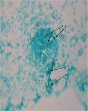
Apoptotic figures appear as small dark spots in this image of developing thyroid in an HH22 embryo that has been counterstained with Fast Green. 1 marks apoptotic bodies in the thyroid and 2 marks clusters of apoptotic bodies in a stran of tissue. The latter suggests that apoptosis is involved in earliest removal of the thyroglossal duct. |
to SAMiller's research
page
to SAMiller's publications
to SAMiller
's homepage


This page was last modified: October
2005
