
similar
appearance of early mouse and
chick embryos
impressive
growth
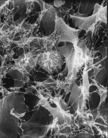
what is it? a
cluster of grapes?
what is it? fallen
leaves?
and purse
strings too?
|
|
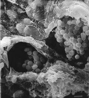
What is it? What may look like clusters of grapes at the mouths of tunnels are actually clusters of embryonic blood cells in the earliest blood vessels. Part of the vascular wall was removed to allow this view in a scanning electon micrograph. We are looking down into these vascular spaces from what is called the extraembryonic coelom.The bumps in the layer that is draped over the top of this picture are the mesoderm cells lining the extraembryonic coelom.The rough broken edge of the yolk sac can be seen at the bottom of this picture. Chick embryo blood islands at 30 hours of incubation enclose clusters of embryonic erythrocytes that will soon begin to circulate in the emerging vascular network. The heart begins to beat at about this time, but blood "islands" are still connecting to each other to form vessels, and the majority of the earliest vessels are technically outside the body in the yolk sac of the new embryo. A careful look might suggest that these blood cells are stuck together, and that is truer than this type of image can show. These blood cells are still connected by gap junctions that allowed synchrony in their early development. Changes in the cell membranes and loss of these junctions would soon have released these blood cells to circulate as individuals in the new chicken. |
|
© 1975 SAMiller |
|
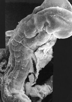
What is it? What might look like a pile of fallen leaves with spider webs is actually cells crawling through a matrix to form the middle layer, the mesoderm, in a chick embryo that has been incubated about 24 hours. The current arrangement of these cells is termed mesenchyme -- loosely associated cells functioning as individuals in an extracellular matrix. There are three basic layers in an early embryo, and each of these layers has a mesenchymal component. Mesectoderm cells come from the neural crest, primordial germ cells migrate from extraembryonic endoderm to the gonad rudiments, and all of the mesoderm moves as mesenchyme at some point in early development. This scanning electron micrograph shows most of the mesenchyme cells as smooth-sided shapes apparently suspended in a web. That "web" is what remained of the extracellular matrix after this chick embryo was fixed, critical point dried and coated with thin layer of metal so that it could be viewed in an electron beam. Migrating cells use this web-matrix to move through embryonic spaces. Notice one cell in the middle that looks round and appears to have blisters. Cell biologists call these blisters blebs. Although blebs can be a sign of an abused or dying cell, they are also often a part of the surface of cells in the phase of the cell cycle called G2, just before the cell divides. We cannot be certain what the condition of this cell was just by looking at its exterior, but the good condition of the rest of the cells in the image suggests that the blebbed cell was not abused in processing. |
© 1975 SAMiller |
|
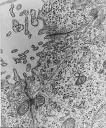
Purse strings? Transmission electron microscopy provides an even closer look within cells of embryos. Images such as these represent a moment of time fixed in place, and this sort of investigation helped inform biologists about some very dynamic activities of cells. In these two images you can see most of one cell, and parts of its two neighbors. The second picture is a larger magnification of part of the first image.These cells are located in a layer called endoderm that will fold to form early gut tube in a mouse embryo. These images show a phenomenon that developmental biologists compare to the pulling of strings to close a purse. In this case the strings are microfilaments that behave like small muscles within cells. You can see these filaments spanning between junctions at the cell membrane. When these filaments shorten (like a contracting muscle) they pull the sides of this end of the cell together. Sections of these cells suggested bottles or flasks, and the term, flask cell, was also applied to this phenomenon. Localized changes in cell shape are a fundamental contributor to much, but not all, of the shaping of early embryos. Formation of large-scale folds does not require assistance at small points like these changes in cell shape; local differences in cell division can produce the same effect. You can simulate this formation by placing your hands on each side of a sheet of paper that is lying on a table top then moving your hands toward each other while pushing down slightly on the paper so that the paper forms a fold and then a tube. Cell shape changes would make a sharper fold, but pressure of growth can also make a fold. Chick embryos do not show cells like these as they fold their endoderm to make a gut tube. Cell shapes in the tightest ectoderm folds suggest that this purse-string phenomenon might be present to help push those two folds toward closure, but we did not find similar images in chick body wall ectoderm. What else can you see in these images? Notice the many circles of cell parts that are adjacent to the narrowed end of this cell. These represent extensions of the cell that were caught in the thin slice used to make this image. A thin slice does not include the attachment of these extensions to the cell. Little folds in the membrane result from compression of the membrane when microfilaments contract, as suggested in these images. Small projections that look like these in section might also be more permanent arrangements on a cell membrane called microvilli. Microvilli increase surface area for absorbtion. They are common on endoderm cells in a yolk sac and later in cells that line absorbtive parts of the adult gut. Microvilli are also abundant on oocytes (egg cells). Finally, there are circular inclusions in these cells near the junctions along the membranes where the microfilaments attach. These circular organelles contain ovals of membrane. The organelles are called mitochondria and can be considered batteries placed near the action so that the energy they produce is close at hand. |
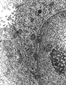 ©
1970 SAMiller ©
1970 SAMiller
A closer view of the
"strings". |
 ©
1970 SAMiller
©
1970 SAMiller