Domains of Differential Cell Proliferation Suggest Hinged Folding in Avian Gut Endoderm

Sue Ann Miller, Michael Adornato, Ailish Briglin, Megan Cavanaugh, Todd Christian, Kelly Jewett, Cheryl Michaelson, Tama Monoson,
Frank Price, Julie Tignor, and Dean Tyrell
Department of Biology, Hamilton College, Clinton, NY

Abstract


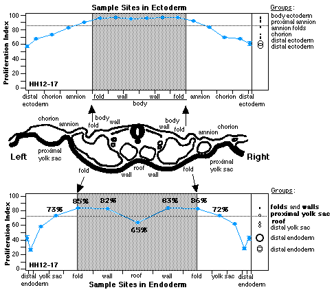
A profile of proliferative growth assessed with tritium autoradiograms from White Leghorn embryo stages Hamburger-Hamilton 6-21 labeled in ovo presents evidence of hinged folding driven by localized differential cell proliferation in endoderm. There is a significant, bilateral pattern, and differences are most pronounced in axial levels that are folding and rotating. Highest proliferation is in cells producing folds; lowest proliferation is in median cells. Localized changes in cell shape are lacking, as are TUNEL markers and cell morphology that would suggest involvement of apoptosis. Folding endoderm to form gut tube appears to be a process that is driven by domains of high cell proliferation flanking a domain of significantly lower proliferation. When considered in the context of an epithelium attached to subjacent mesoderm, these differentials could produce a forced and directed buckling of endoderm into lateral folds that join and enclose a tube. Patterns suggest that endoderm folds about a median hinge. In the light of this new information, we suggest it is more precise to refine the term, median hinge point (MHP), to neural hinge point (NHP) and gut hinge point (GHP).

Grant sponsor: Howard Hughes Medical Institute; Grant sponsor: Margaret Bundy Scott Fellowship; Grant sponsor: Casstevens Family Fund; Grant sponsor: Hamilton College Faculty Research Funds; Grant sponsor: Hamilton College Academic Fund for Seniors.

Developmental Dynamics 216:398-410 (1999)

© 1990 SAMiller


The proliferative hinge in median endoderm.

Data taken from comparable sites and axial levels within the same chick embryos show different profiles of proliferation during folding of these membranes. An enlarged tracing of a representative section shows placement of sample sites and helps to place the statistics into morphological perspective. Gray in the data graphs indicates areas between body folds in each layer as clarified by arrrows from the tracing. Space between each pair of arrows represents 1 mm. Data combined from 6 folding levels show a typical pattern for folding endoderm with gut roof as a significantly separate domain within body endoderm. The summary of composite data from ectoderm was published previously (Miller, et al. 1994) and is included in this figure for comparison of the two germ layers. Connected means were derived using ANOVA. Comparison circles were derived by testing each pair with the student's t test and show significance at the 0.05 level. Size of comparison circles is informative: a smaller circle indicates greater the sample size and confidence in the mean it represents. Circles for means that are significantly different either do not intersect or intersect slightly so that the outside angle of intersection is less than 90°. Significantly different sets of data identified as "groups" were confirmed while running the JMP program. (n=39, stages HH 12-17; 15- to 30-somite pairs)


There is similar evidence of a hinge in mouse endoderm.
A preliminary report of data from both chick and mouse embryos was presented at Experimental Biology 99.

to SAMiller's research page
to SAMiller's publications
to SAMiller's homepage

Last Modified: 9 December 1999