Cell Proliferation in Chick Oral Membrane
Lags Behind that of Adjacent Epithelia at the Time of
Rupture
Sue Ann Miller and Christopher W. Olcott
Department of Biology, Hamilton College, Clinton, New York
Abstract
Radioautographic analysis showed that ectoderm and endoderm cells
in chick oral membrane continued to label with tritiated thymidine
through the period of rupture, but their frequency of labeling was
significantly lower than those of adjacent epithelia. Frequency of
labeling increased in adjacent ectoderm and endoderm, while oral
membrane rates remained relatively low, suggesting that growth in the
membrane lags relative to adjacent epithelia. Relatively greater
proliferation in adjacent epithelia could generate tension and pull
apart the thinned oral membrane. Differentials in rates of cell
proliferation, when considered along with knowledge of cellular
rearrangements following changes in basal lamina and matrical
components, suggest that differential growth is an important force in
rupture of the avian oral membrane.
Anatomical Record 223:204-208 (1989)
|
|
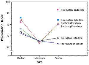
Figure 5 Graphic display of average proliferation
indices at five sites in 11 embryos shows differentials that
occurred when adjacent epithelia increased rate of labeling
while oral membrane cells lagged at lower frequencies of
labeling.
|
|
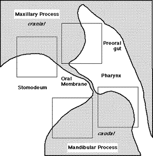
Figure 1 Typical sample sites around the oral
membrane in a sagittal section. Boxes represent placement of
the ocular grid over ectoderm on the maxillary and
mandibular processes (stomodeum) and endoderm in the preoral
gut and pharynx. The entire oral membrane consituted one
site.
|
|
back to research
back to publications
return to Professor SAMiller
's homepage


Last Modified: 3 October
1999