Apoptosis Removes Chick Embryo Tail Gut
and Remnant of the Primitive Streak
Sue Ann Miller and Ailish Briglin
Department of Biology, Hamilton College, Clinton, NY
Abstract
Developmental Dynamics 206:212-218 (1996)
Removal of transient features in morphogenesis of chick embryo
tail is by programmed cell death. We used ApopTag™
(Oncor®, Gaithersburg, MD) with the peroxidase/DAB
procedure to correlate apoptosis with earlier reports of patterns of
cell death in stage HH17-25 embryos, and our results suggest that the
cell death inferred with supravital staining and appearance of cells
in morphogenesis of the tail bud is programmed cell death called
apoptosis.
Apoptosis markers in tail bud are most abundant in the median cell
cord of occluded, degenerating tail gut. Tail bud mesenchyme marks
for apoptosis most frequently in the ventrum of older stages, where
cell death has been reported. Cells of the remnant of the primitive
streak (Hensen's Node) mark for apoptosis, suggesting that programmed
cell death is a stop signal for axial organization at the caudal
terminus. Apoptosis markers in postmembrane cloacal endoderm
anticipate the transient cloacal fenestra. Lack of apoptosis markers
in neural tube, notochord, and somites supports the suggestion of
Schoenwolf (1981) that cells of those areas in the tail bud are
assimilated into the growing rump of the chick embryo. Lack of
markers in neural tube of tail bud formed by secondary neurulation
suggests that apoptosis is not involved in cavitation of medullary
cord, but further investigation is necessary.
A limited investigation of pharyngeal membranes and midgut, where
cell death has not been reported as important in morphogenesis, did
not show apoptosis markers in those tissues (Miller and Briglin,
1994). Absence of apoptosis markers in roof of gut tube suggests that
the lower frequency of thymidine labeling reported for those cells
(Miller, 1986) is not a result of apoptosis. Clearly marked cells
correlate with expected locations of migrating neural crest and
primordial germ cells in these stages, but distribution of apoptosis
markers was not abundant or general for either cell type.
[Note: Color prints are in the publication, but black
and white may show details better in this format. I installed both
color and a black and white versions of each figure until I have more
time to work on my scanning skills and to decide which is most
effective in this presentation.]
|
|
|
|
|
|
|
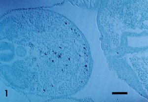
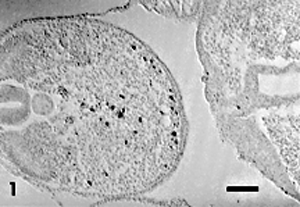
Figure 1. Stage 21; tail bud and
cloaca. Brown apoptosis markers are concentrated along
median site of degenerating tail gut and in ventral tail bud
mesenchyme near the site of the remnant of the primitive
streak. Cloacal plate does not mark for apoptosis. Bar = 100
µm
|
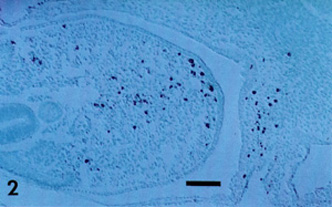
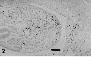
Figure 2. Stage 22; tail bud and
cloacal area. Brown apoptosis markers are more frequent in
concentration at the site of degenerating tail gut and
remnant of primitive streak. Mesenchyme in proximal and
ventral tail bud adjacent to cloacal plate shows frequent
markers of apoptosis. Bar = 100 µm
|
Grant sponsor: Howard Hughes Medical Research
Foundation; Grant sponsor: Sigma Xi; Grant sponsor: The Casstevens
Family Fund; Grant sponsor: Hamilton College Faculty Research Funds;
Grant sponsor: The Hamilton College Academic Fund for
Seniors.
to SAMiller's research page
to SAMiller's publications page
to SAMiller 's homepage


Last Modified: 21 September
1999